Attic Cholesteatoma Radiology

Fd Acquired Pars Flaccida Cholesteatoma Left Coronal T Bone Ct Image Shows An Atticoantral Nondependent Homogeneous Soft Radiology Image Shows Head And Neck

Cholesteatoma

Acquired Cholesteatoma Radiology Reference Article Radiopaedia Org

Mastoditis Middle Ear Head And Neck Sinusitis

Choleastoma In 2020 Middle Ear Eustachian Tube Dysfunction Ear Infection

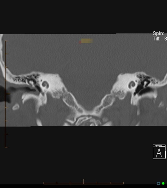
Cholesteatoma Radiology Reference Article Radiopaedia Org
The external acoustic canal is a rare location for a cholesteatoma with an estimated incidence around 1 2 per 1 000 new otological patients.
Attic cholesteatoma radiology.

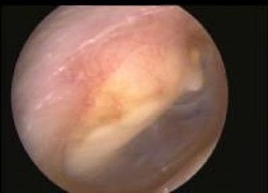
Eardrums Seen In 8 Conditions Normal Eardrum Acute Otitis Media Perforation Small Perforation Attic Perforat Otitis Otitis Media Health Assessment Nursing
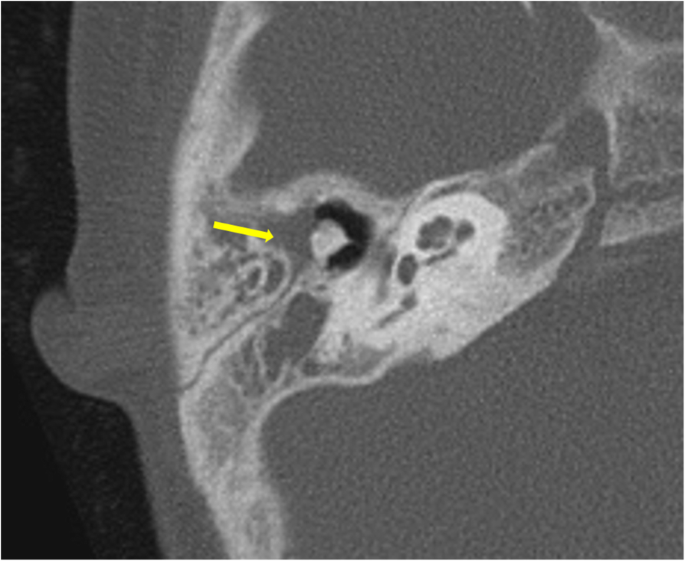
Hrct Imaging Of Acquired Cholesteatoma A Pictorial Review Springerlink
Pars Tensa Cholesteatoma Radiology Case Radiopaedia Org

Cholesteatoma Radiology Case Radiopaedia Org
Source : pinterest.com
